Retinal detachment is a sight-threatening emergency, and high myopia significantly raises the risk. Here's what to watch for, why it happens, and what urgent treatment involves.
The retina is a thin layer of light-sensitive tissue lining the back of the eye, and it depends on the tissue directly beneath it for its blood supply and nourishment. Retinal detachment happens when the retina physically separates from that underlying layer, cutting off its supply and, if untreated, causing permanent loss of function in the affected area.
Unlike most of the conditions we cover on this site, retinal detachment isn't something to monitor over months or years. It's a sight-threatening emergency that typically develops from a retinal tear, and the window for effective treatment is measured in hours to days, not weeks. The earlier it's caught and treated, the better the chances of preserving vision.
This is also a condition we think about constantly in the context of myopia management. High myopia is one of the strongest risk factors for retinal detachment, which is a significant part of why we take slowing myopia progression in children so seriously.
Retinal detachment usually gives some warning before vision is seriously affected. Knowing these signs, and acting on them quickly, is genuinely sight-saving.
A sudden increase in floaters, especially many appearing at once, is different from the occasional floater most people have long-term.
Brief flashes or streaks of light, particularly noticeable in peripheral vision or in dim lighting, can signal the retina being pulled or torn.
A dark shadow or curtain effect moving across part of your vision, often starting at the edges, is one of the clearest signs of an active detachment.
A loss of side vision, sometimes described as looking through a narrowing tunnel, can develop as the detachment spreads.
If the detachment reaches the macula, central vision can blur or drop quickly, which is a sign the situation has become more urgent.
New floaters and flashes are common and usually harmless, but if they arrive suddenly or worsen quickly, treat it as urgent and get assessed the same day.
In a highly myopic eye, the eyeball is longer than average, back to front. That extra length doesn't come for free: it stretches the retina thinner, particularly towards its outer edges, and makes the vitreous gel inside the eye more likely to pull away unevenly as it changes with age. Both of these increase the chance of a retinal tear developing, which can progress to a full detachment if it isn't caught.
This is one of the clearest, most direct links between childhood myopia and lifelong eye health, and it's a large part of why we focus so heavily on slowing myopia progression rather than simply correcting it with stronger glasses each year. Every dioptre of myopia we can prevent in childhood measurably reduces the lifetime risk of retinal detachment, along with other myopia-related eye diseases.
Other risk factors include a previous retinal detachment in the other eye, a family history of detachment, previous eye surgery or significant trauma, and lattice degeneration, a specific pattern of retinal thinning we look for during a dilated examination.
Most retinal detachments begin the same way: the vitreous, a clear gel filling the back of the eye, gradually shrinks and separates from the retina as part of a normal age-related process called posterior vitreous detachment. Usually this happens without incident. Occasionally, though, the vitreous stays firmly attached to the retina in one spot and, as it pulls away, tears a small hole in the retinal tissue.
Once a tear exists, fluid from inside the eye can pass through it and seep beneath the retina, gradually lifting it away from the tissue that supports it, which is what we mean by a detachment. Where and how quickly this spreads determines how urgently it needs to be treated, particularly whether it threatens the macula, the area responsible for central vision.
Because a highly myopic retina is thinner and under more mechanical stress to begin with, both the initial tear and the progression to detachment tend to happen more readily than in an eye of typical length.
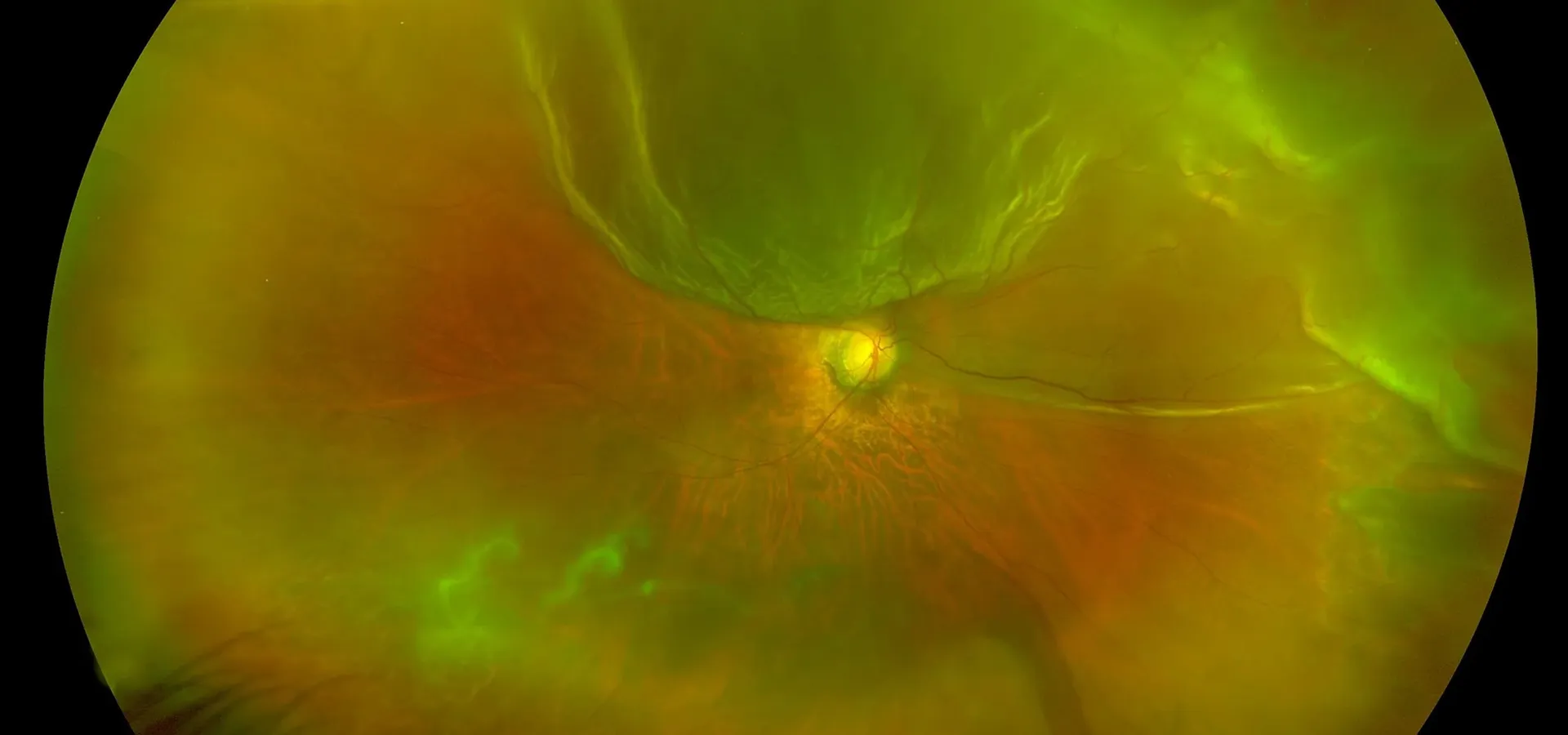
Diagnosing a retinal tear or detachment relies on getting a clear, detailed view of the peripheral retina, which is why a dilated examination is central to this assessment.
Widening the pupil gives us a full view right to the edges of the retina, where tears and early detachment most often begin.
Ultra-widefield photography documents the peripheral retina in detail, useful both for diagnosis and for tracking known areas of thinning over time.
Where detachment is suspected near the macula, OCT confirms whether it's involved, which materially affects both urgency and expected outcome.
Mapping which areas of vision are affected helps confirm the extent of a detachment and correlates it with what we see on examination.
If you notice a sudden shower of floaters, new flashes of light, or a shadow moving across your vision, call us and tell us it's urgent. We prioritise same-day assessment for these symptoms, because the outcome of retinal detachment treatment is genuinely time-sensitive. If we can't see you immediately, we'll help direct you to emergency eye care.
Unlike most eye conditions, this isn't a pathway of gradual monitoring. Once a tear or detachment is confirmed, the priority is fast, coordinated action.
If you present with warning signs, we prioritise a dilated examination that same day rather than at a routine follow-up appointment.
Dilated examination, wide-field imaging and OCT together confirm whether there's a tear, a detachment, or both, and how far it extends.
Confirmed tears and detachments are referred directly to a retinal surgeon, usually the same day, given how much timing affects the outcome.
Depending on the type and extent, treatment may involve pneumatic retinopexy, a scleral buckle, or a vitrectomy, each aiming to reattach the retina and seal the original tear.
Recovery is closely monitored by the treating surgeon, with follow-up examinations to confirm the retina remains attached and healing as expected.
Once treated, we continue monitoring both eyes long term, since having one retinal detachment raises the risk of a future tear, particularly in a fellow highly myopic eye.
Retinal detachment risk is a thread that runs through a lot of what we do, not just as an emergency response but as a reason we take myopia management so seriously from childhood. Reducing how myopic a child becomes measurably reduces their lifetime risk of retinal complications, and it's one of the most concrete, evidence-based reasons we recommend myopia control treatment rather than simply updating glasses each year.
For anyone with high myopia already, we build regular dilated retinal examinations and wide-field imaging into routine care, so thinning or early changes can be identified before they become a tear. And if you ever notice sudden floaters or flashes, we treat that as urgent, not something to mention at your next scheduled visit.
Our Kew East practice is easily accessible from across Melbourne's inner east, including Hawthorn, Camberwell, Balwyn and Box Hill.
If you're experiencing sudden floaters, flashes or a shadow in your vision, please call us directly rather than booking online. For everyone else, particularly if you have high myopia, a comprehensive dilated assessment is the best way to understand your retinal health.