Macular assessment for age-related macular degeneration at Eyecare Concepts Melbourne
Eye Health Conditions

Macular Degeneration: Protecting Your Central Vision

AMD affects the macula, the part of the eye responsible for the sharp central vision you rely on to read, drive and recognise faces. Here's how it develops, how we monitor it, and what treatment involves.

What Is Macular Degeneration?

The macula is a small area at the centre of the retina, only a few millimetres across, yet it's responsible for the sharp, detailed vision you use for almost everything that matters day to day: reading a menu, recognising a face across the room, threading a needle, or reading a number plate. Everything around the edges of your vision is handled by the rest of the retina, but that fine central detail comes from the macula alone.

Age-related macular degeneration, or AMD, is a progressive condition affecting this area. It develops in two forms. Dry AMD, the more common type, involves a gradual thinning of macular tissue and a build-up of small deposits called drusen. Wet AMD is less common but more urgent, involving abnormal blood vessels growing beneath the macula that can leak fluid or blood.

Because the macula handles central vision specifically, AMD doesn't cause total blindness. Peripheral vision is preserved, but the tasks that rely on fine central detail become progressively harder, which is why early detection and monitoring matter so much.

macular-degeneration-02.jpg — Macula Anatomy Photo
Recognising The Signs

How AMD Affects Your Vision

Because AMD affects central vision specifically, the signs often show up in fine detail tasks before anything else changes. Here's what to watch for.

Distortion On A Grid

Straight lines can appear wavy or bent, most easily noticed using an Amsler grid, a simple pattern of straight lines used to detect this early.

Blurred Or Blank Central Patch

A hazy or missing area can appear in the centre of vision, while the surrounding field stays clear.

Difficulty Recognising Faces

Fine facial detail relies heavily on central vision, so faces can become harder to recognise, particularly from a distance.

Needing More Light To Read

Reading and close work can start requiring brighter, more direct light than it used to.

Colours Appearing Less Vivid

Colours can look duller or less saturated than before, a subtle change that's easy to attribute to other things.

Sudden Distortion Or Vision Loss

A rapid change, particularly new distortion or a sudden drop in central vision, can signal wet AMD and needs urgent review.

macular-degeneration-03.jpg — Amsler Grid Self-Test Photo

Why Macular Health Deserves Attention

AMD is one of the leading causes of vision impairment in people over 50, and it targets exactly the vision most people rely on most: reading, driving, screens, and faces. Because it doesn't affect peripheral vision, someone can technically still see, while finding the everyday tasks that depend on fine detail increasingly frustrating.

Age is the single biggest risk factor, but it isn't the only one. A family history of AMD raises individual risk meaningfully, smoking roughly doubles it, and high myopia carries its own related risk, a distinct process called myopic maculopathy, where the physical stretching of a highly myopic eye thins and stresses the macula in a similar way.

Wet AMD, in particular, can progress over days to weeks rather than years, which is why self-monitoring at home between visits, alongside regular comprehensive assessment, genuinely changes outcomes.
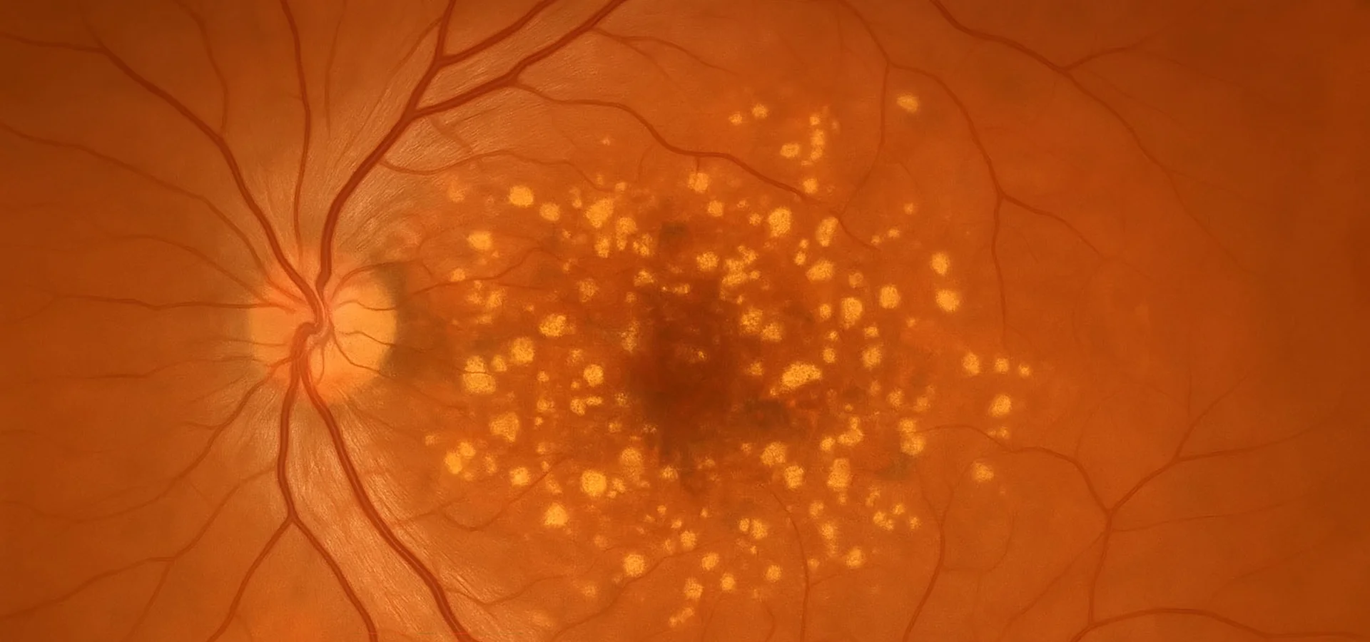
The Science Of Dry And Wet AMD

The macula sits on a layer called the retinal pigment epithelium, which supports and nourishes the light-sensitive photoreceptor cells above it. In dry AMD, waste products accumulate beneath the retina as small yellow deposits called drusen, and over time this layer thins and photoreceptors are gradually lost, a process called geographic atrophy in its more advanced form. Dry AMD accounts for the large majority of cases and usually progresses slowly, over years.

Wet AMD develops when the body attempts to grow new blood vessels beneath the macula in response to stress or lack of oxygen, driven by a signalling protein called VEGF. These new vessels are fragile and prone to leaking fluid or blood, which distorts and damages the macula far more quickly than the dry form. Wet AMD can develop from existing dry AMD or, less commonly, appear as the first sign of the condition.

Understanding which form, and which stage, is present is what allows us to recommend the right monitoring interval and, where relevant, the right treatment pathway.

macular-degeneration-04.jpg — Retinal Layer Diagram
Detection & Diagnosis

How We Detect And Diagnose AMD

Diagnosing AMD accurately, and catching change early, relies on a combination of functional testing and detailed imaging of the macula itself.

Amsler Grid Testing

A simple grid of straight lines checked one eye at a time, used to detect distortion or blank areas in central vision.

OCT & Retinal Imaging

Cross-sectional scans reveal the individual retinal layers beneath the macula, picking up fluid, drusen or thinning long before symptoms appear.

Central Visual Field Testing

Maps the sensitivity of central vision specifically, identifying subtle blind spots that broader tests can miss.

Dilated Macular Examination

A direct view of the macula itself, checking for drusen, pigment changes or signs of fluid consistent with wet AMD.

Self-Monitoring At Home

Between visits, checking an Amsler grid at home, one eye at a time, is one of the most useful things someone with AMD or at higher risk can do. Because wet AMD can progress over days rather than years, catching new distortion or a new blank patch early and having it reviewed promptly can genuinely change the outcome. We provide an Amsler grid and show you exactly how to use it as part of your assessment.

Management Options

Macular Degeneration: What Actually Helps

Glasses don't correct AMD, since it isn't a focusing problem. Management depends on which form is present and how advanced it is, ranging from monitoring and nutrition through to urgent treatment for wet AMD. Here's how that pathway typically unfolds.

1

Comprehensive Assessment & Baseline Imaging

OCT and retinal imaging establish a detailed baseline of your macula, so any future change can be tracked accurately against your own starting point.

2

Risk Assessment & Home Monitoring

We discuss your individual risk factors and set up Amsler grid self-monitoring, so change between visits is more likely to be caught early.

3

Lifestyle & Nutritional Support

Not smoking, eating a diet rich in leafy greens, and where appropriate, AREDS2 supplementation for those at intermediate or advanced risk of dry AMD progression.

4

Regular Monitoring Reviews

Scheduled OCT reviews track subtle change in the retinal layers over time, adjusting the monitoring interval based on what we find.

5

Urgent Referral For Wet AMD

Where imaging or symptoms suggest wet AMD, we arrange prompt referral to an ophthalmologist for anti-VEGF injections, which can stabilise or improve vision when started early.

6

Ongoing Co-Managed Care

Whether dry or wet, AMD is managed long term. We stay involved alongside any treating ophthalmologist, coordinating your monitoring and reviews.

macular-degeneration-05.jpg — Clinic Consultation Photo

Our Approach To Macular Care

Because the macula is small and change can be subtle, our focus at Eyecare Concepts is on precise, repeatable OCT imaging that lets us compare your retinal layers visit to visit with real confidence, rather than relying on how vision feels alone.

We also take family history and lifestyle factors, including smoking and high myopia, seriously when setting your monitoring schedule, and talk through whether AREDS2 supplementation is appropriate for your individual risk, rather than recommending it as a blanket approach. Where wet AMD is suspected, we prioritise fast referral, given how much timing matters to outcomes.

Our Kew East practice is easily accessible from across Melbourne's inner east, including Hawthorn, Camberwell, Balwyn and Box Hill.

macular-degeneration-06.jpg — Macular Degeneration Review Photo
Common Questions

Macular Degeneration FAQs

What is the difference between dry and wet AMD?
Dry AMD is more common and involves gradual thinning of the macula and a build-up of deposits called drusen, usually progressing slowly over years. Wet AMD is less common but more urgent, involving abnormal blood vessels growing beneath the macula that can leak fluid or blood and cause faster vision loss.
What age does macular degeneration start?
AMD becomes more common from age 50 onwards, with risk increasing steadily with age. Early changes are often picked up during a comprehensive eye exam years before any symptoms appear.
What are the first signs of macular degeneration?
Early signs can include mild blurring of central vision, difficulty reading in low light, or straight lines appearing slightly wavy on an Amsler grid. Many people notice no symptoms at all in the earliest stages, which is why regular screening matters.
Can macular degeneration be cured?
No, but it can be managed. Dry AMD progression can often be slowed through monitoring, lifestyle changes and, for those at higher risk, AREDS2 supplementation. Wet AMD is treated with anti-VEGF injections, which can stabilise or improve vision, though it isn't a permanent cure.
Will macular degeneration make me completely blind?
No. AMD affects central vision but doesn't cause total blindness, since peripheral vision is preserved. This is why people with AMD can usually still navigate their environment independently, even as central tasks like reading and recognising faces become harder.
Can diet help prevent macular degeneration?
A diet rich in leafy greens and fish, combined with not smoking, is associated with lower AMD risk. For those already at intermediate or advanced risk, specific AREDS2 supplementation may also help, though this should be guided by an individual assessment rather than assumed.
Is macular degeneration hereditary?
It can run in families. Having a parent or sibling with AMD raises your own risk, which is one of several factors, alongside age, smoking and high myopia, that we consider when assessing your individual risk.
Should I take eye vitamins if I have a family history of AMD?
Not automatically. AREDS2 supplements have been shown to help specifically at intermediate or advanced stages of dry AMD, not as a general preventative measure for everyone with a family history. We assess your individual macular health before recommending them.
How often should I check my Amsler grid at home?
If you've been diagnosed with AMD or are at higher risk, checking one eye at a time, roughly once a week, is a reasonable habit. Any new distortion, wavy lines or blank patch warrants a prompt review, since wet AMD can progress quickly.
Is myopic maculopathy the same as age-related macular degeneration?
No, though they can look similar on imaging. Myopic maculopathy is driven by the physical stretching of the eye in high myopia, while AMD is age-related. Both affect the macula and are monitored with similar OCT and retinal imaging techniques.

Book A Comprehensive Eye Assessment

Macular changes are easiest to manage when caught early. A comprehensive assessment with OCT imaging gives us a clear picture of your macular health and, where needed, the most appropriate monitoring or treatment plan for you.

Book an Appointment