Glaucoma damages the optic nerve, usually without any warning signs in its early stages. Here's how it develops, why regular testing matters more than symptoms, and how it's managed.
Glaucoma is often called the silent thief of sight, and for good reason. In its most common form, it causes no pain, no blurring and no obvious warning in its early stages. By the time someone notices a change in their vision, the condition has usually already been progressing for years.
Glaucoma describes a group of conditions that damage the optic nerve, the bundle of over a million nerve fibres that carries visual information from the eye to the brain. This damage is most often linked to pressure building up inside the eye, though glaucoma can also occur when eye pressure is within a normal range. As nerve fibres are lost, so is the corresponding area of vision, usually starting at the outer edges of sight.
Because this early vision loss happens at the periphery, where it's easiest for the brain to compensate for, most people don't notice anything is wrong until a significant portion of the optic nerve has already been affected. This is precisely why glaucoma is diagnosed through testing, not symptoms, and why regular comprehensive eye examinations matter so much, particularly for those with a family history.
The most common form of glaucoma produces no symptoms at all until vision loss is advanced. Where signs do appear, whether gradually or, in rarer cases, suddenly, these are the changes to be aware of.
The most common type progresses silently for years. Central vision stays sharp while peripheral vision is quietly lost.
Side vision narrows slowly, often described only in hindsight as bumping into things or missing objects to the side.
Small gaps can appear in the field of vision, which the brain often masks until they overlap or grow larger.
In advanced, untreated cases, only a narrow, central field of vision remains, as though looking through a tube.
Rings of light around lamps or headlights can appear, particularly in the lead-up to an acute pressure spike.
A rapid rise in eye pressure can cause severe eye pain, headache, nausea and blurred vision, requiring urgent care.
Glaucoma is a leading cause of irreversible blindness worldwide, and the vision it takes doesn't come back. Nerve fibres, once lost, cannot be regenerated, which makes early detection and consistent management the only real defence.
Because early loss affects peripheral vision first, the impact on daily life often shows up gradually, in missed obstacles, difficulty with steps and kerbs, or a slower reaction to hazards while driving, long before someone would describe their vision as blurry. Left unmanaged, this narrowing can eventually threaten independence and central vision itself.
Family history matters here more than with many other eye conditions. Having a parent or sibling with glaucoma meaningfully raises your own risk, alongside factors like high myopia, diabetes and age. A comprehensive assessment, rather than a basic vision check, is what actually detects it.
Inside the eye, a clear fluid called aqueous humour is constantly produced and drained, maintaining a stable internal pressure. This fluid drains through a mesh-like structure called the trabecular meshwork, at the angle where the iris meets the cornea. When drainage slows or becomes blocked, pressure inside the eye rises, and that pressure can gradually damage the delicate nerve fibres at the back of the eye.
The most common form, primary open-angle glaucoma, develops slowly as the drainage angle remains open but becomes less efficient over time, often with no symptoms until significant nerve damage has occurred. Angle-closure glaucoma is less common but more urgent, occurring when the drainage angle becomes physically blocked, sometimes causing a sudden, painful spike in pressure that needs emergency treatment. A third pattern, normal-tension glaucoma, causes the same nerve damage despite eye pressure staying within a typical range, which is why assessing the optic nerve directly matters as much as measuring pressure.
Understanding which pattern is present, and how the optic nerve and visual field are responding, is what allows treatment to be targeted rather than generic.
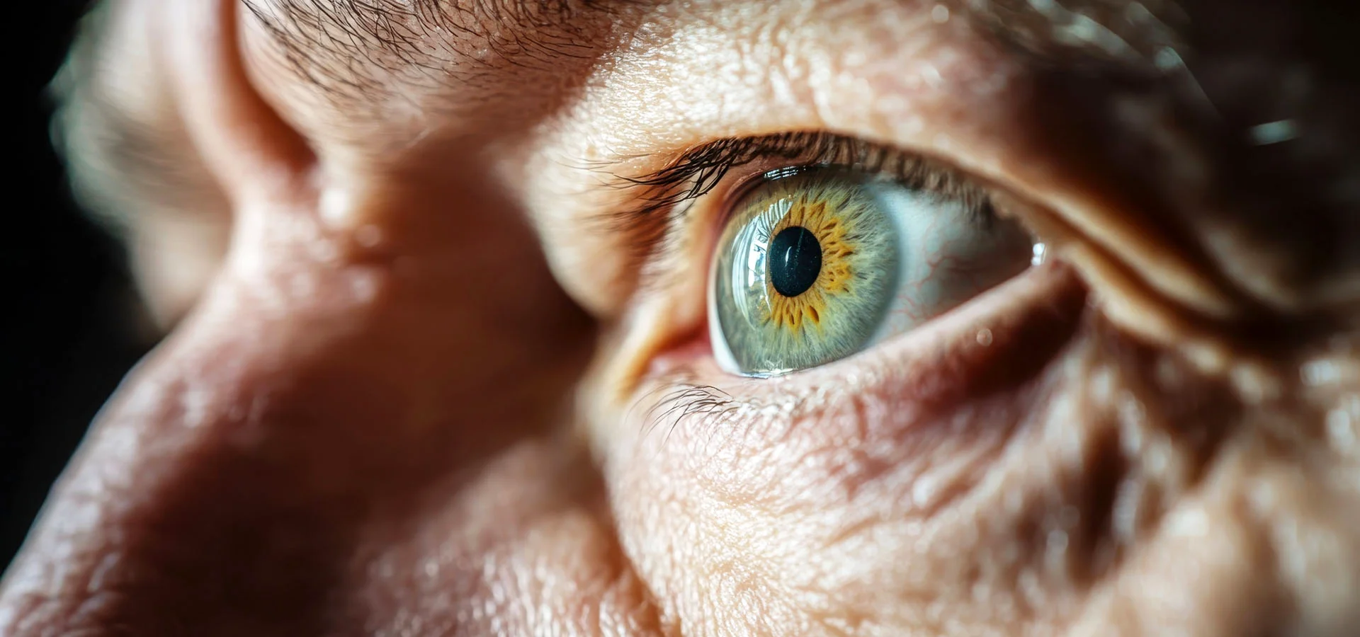
Because glaucoma rarely announces itself, diagnosis relies on a set of specific tests performed during a comprehensive eye examination, not on how well you feel you're seeing.
Measures the pressure inside the eye, a key risk indicator and one of the first things we check.
Optical coherence tomography (OCT) measures the thickness of the nerve fibre layer at the back of the eye, giving us a precise structural baseline we can compare against on every future visit.
Maps your full field of vision to identify blind spots or narrowing that you wouldn't otherwise notice.
Examines how fluid drains from the eye, distinguishing open-angle from angle-closure patterns of glaucoma.
Visual field testing measures how glaucoma affects your sight, but it has a blind spot of its own: by the time a visual field defect shows up, research suggests more than half of the relevant nerve fibres may already be lost. OCT looks at the structure behind the function, measuring nerve fibre layer thickness directly and picking up thinning years before it would ever appear as a gap in your visual field.
Because OCT produces a precise, repeatable measurement, we can track subtle change from one visit to the next against your own baseline, rather than waiting for symptoms or a functional test to catch up. This is why we recommend OCT scanning as part of routine eye care for everyone over 40, and earlier still for anyone with a family history of glaucoma.
OCT is included as standard in our Advanced Eye Care and Ultimate Eye Health+ examination packages, so this level of detail is part of a comprehensive assessment, not an optional add-on.
Glasses have no role in treating glaucoma. It isn't a focusing problem, so no lens can correct it. Managing glaucoma is about protecting the optic nerve function you still have, and that starts with lowering eye pressure and continues with lifelong monitoring. Here's how that pathway typically unfolds.
Once glaucoma is suspected, we establish a detailed baseline: eye pressure, optic nerve imaging and visual field results, to measure any future change against.
Regular reviews track your optic nerve and visual field over time. Because glaucoma often produces no symptoms, this monitoring schedule is how progression is actually caught.
We talk through your individual risk factors, family history, eye pressure and nerve findings, so the treatment approach reflects your circumstances rather than a one-size-fits-all rule.
Where treatment is needed, daily eye drops are usually the first step, reducing eye pressure to slow or halt further optic nerve damage.
When drops aren't enough on their own, we refer you to a trusted ophthalmologist to discuss laser treatment or surgery, options that further improve drainage and lower pressure.
Glaucoma is managed, not cured, so care continues indefinitely. We stay involved alongside your ophthalmologist, coordinating your ongoing monitoring and treatment.
Because glaucoma is a lifelong condition, our focus at Eyecare Concepts is on building an accurate, individual picture from the outset and tracking it consistently over time, rather than relying on a single test result in isolation.
Advanced OCT imaging gives us a precise, repeatable structural baseline, so genuine change can be distinguished from natural variation between visits, often years before it would show up in a visual field test. Where treatment or a specialist opinion is needed, we work alongside trusted Melbourne ophthalmologists and remain part of your ongoing care, so monitoring, treatment and reviews stay coordinated rather than fragmented.
We also pay close attention to family history, given how much it can shift individual risk, and encourage relatives of glaucoma patients to have their own comprehensive assessment. Our Kew East practice is easily accessible from across Melbourne's inner east, including Hawthorn, Camberwell, Balwyn and Box Hill.
Glaucoma is detected through testing, not symptoms. A comprehensive assessment gives us a clear picture of your optic nerve health and, where needed, the most appropriate management plan for you.