Diabetes can quietly damage the blood vessels in your retina long before you'd notice any change in vision. Here's how it happens, how we screen for it, and how we work with your GP to protect your eyesight.
The retina relies on a fine network of tiny blood vessels to stay nourished and functioning. Diabetes, when blood sugar runs high over months and years, gradually damages these vessels, causing them to weaken, leak or become blocked. This damage is what we call diabetic retinopathy, and it can affect anyone with type 1 or type 2 diabetes.
What makes diabetic retinopathy particularly important to screen for is that it typically causes no symptoms in its earlier stages. Vision can remain completely normal while changes are already happening at the back of the eye, which is why regular screening matters regardless of how well your diabetes feels managed day to day.
Left unchecked, diabetic retinopathy can progress to more advanced stages, including diabetic macular edema, where fluid builds up in the macula and affects central vision, and proliferative retinopathy, where fragile new blood vessels grow abnormally and put vision at greater risk.
Early diabetic retinopathy usually produces no symptoms at all. Where signs do appear, they often indicate the condition has already progressed, which is exactly why we don't rely on symptoms to catch it.
Vision can stay sharp and comfortable while retinal blood vessel damage is already underway, sometimes for years.
Vision that varies from day to day can relate to blood sugar swings affecting the lens and retina, not just retinopathy itself.
Bleeding from fragile new vessels in more advanced disease can appear as sudden floaters or dark patches.
Colours can appear less distinct or harder to differentiate as the condition affects the retina's function.
Diabetic macular edema, fluid build-up at the macula, can cause blurred or distorted central vision.
In advanced, proliferative disease, bleeding into the eye can cause a sudden, significant drop in vision needing urgent care.
Diabetic retinopathy is one of the leading causes of vision loss in working-age adults, yet much of that vision loss is genuinely preventable with regular screening and timely treatment. The challenge is that by the time symptoms appear, meaningful damage has often already occurred.
Screening isn't just about the eyes in isolation. What we find during a diabetic eye check can be a useful, early indicator of how well blood sugar is being managed overall, which is part of why we place real value on working alongside your GP rather than treating your eye health as a separate, disconnected appointment.
Risk increases the longer you've had diabetes, and with less controlled blood sugar and blood pressure, but regular screening matters at every stage, including for anyone newly diagnosed.
Persistently high blood sugar gradually damages the walls of the retina's smallest blood vessels. Early on, this shows up as microaneurysms, tiny bulges in the vessel wall, and small areas of leakage, a stage known as non-proliferative diabetic retinopathy. At this point vision is usually still unaffected, but the changes are visible on retinal imaging.
As damage progresses, blood vessels can become blocked, starving areas of the retina of oxygen. In response, the eye attempts to grow new blood vessels to compensate, but these new vessels are fragile, poorly formed and prone to bleeding, a more advanced stage called proliferative diabetic retinopathy. Separately, leaking fluid can accumulate specifically at the macula, causing diabetic macular edema, which affects central vision directly.
Understanding exactly which stage is present, and whether the macula is involved, is what determines the right monitoring interval and whether active treatment is needed.
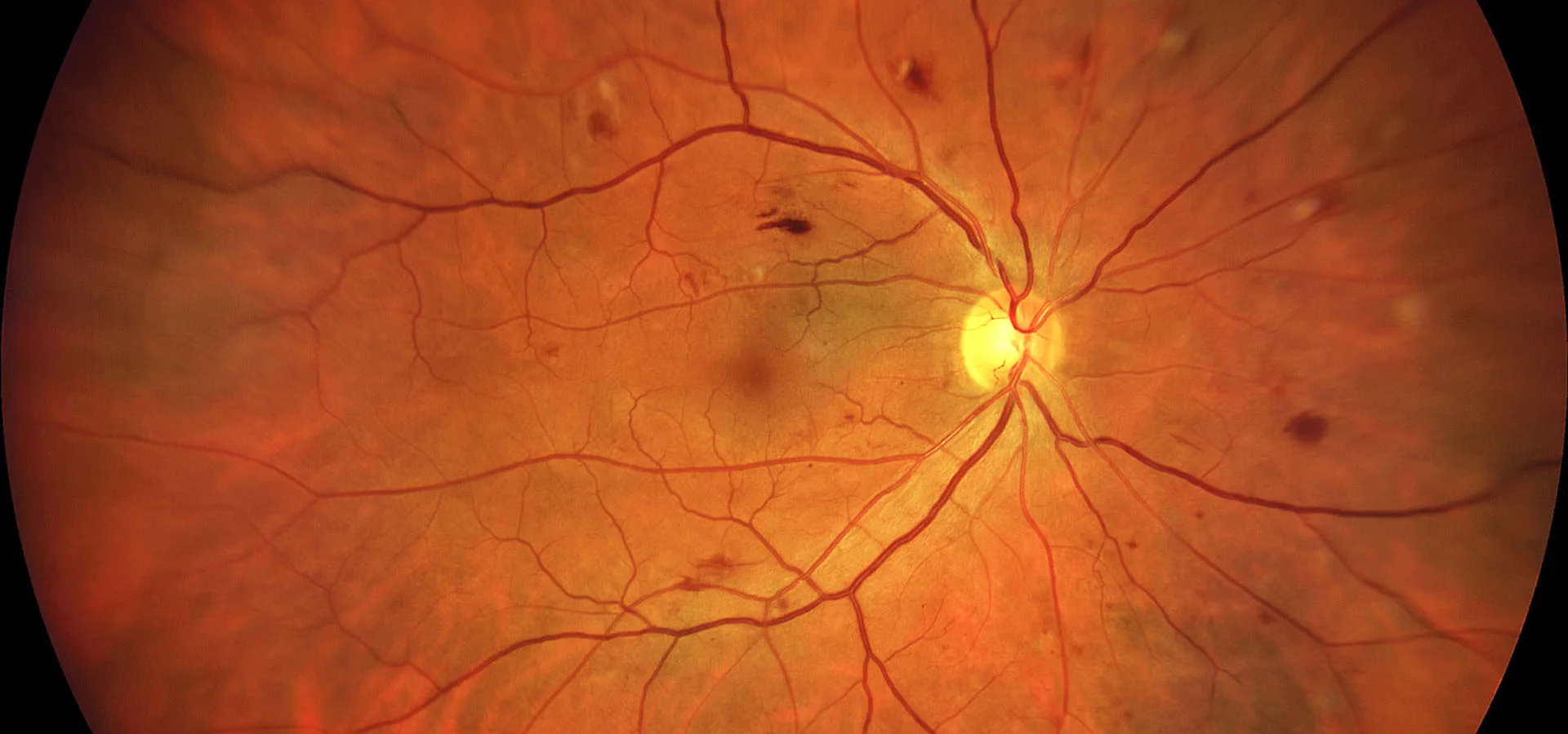
Because early diabetic retinopathy has no symptoms, diagnosis relies entirely on a thorough look at the retina itself, not on how your vision feels.
A single scan captures around 200 degrees of the retina, giving us a detailed, high-resolution view to check for microaneurysms, leakage or new vessel growth.
Diabetic eye checks are dilated as standard, since diabetic changes can appear right at the retina's periphery and are best assessed with a fully widened pupil.
Cross-sectional imaging checks specifically for diabetic macular edema, measuring retinal thickness with precision no photograph alone can match.
We review your vision, diabetes history and recent blood sugar control together, to put what we see on imaging in proper context.
Unlike some of our other assessments, where Optos imaging alone is often enough, diabetic eye checks are generally dilated as a matter of course. Diabetic changes can develop right out at the periphery of the retina, and dilation gives us the most reliable, complete view to make sure nothing is missed.
Because of this, expect your vision to be blurred and light-sensitive for several hours after your appointment. We'd recommend arranging a driver, or allowing time to wait it out before driving yourself home.
Managing diabetic retinopathy is a genuine team effort between you, us and your GP. Here's how that typically comes together.
Optos imaging, a dilated examination and OCT together establish a clear picture of your current retinal health.
With your consent, we ask for your GP's details and send them a report on your eye health findings, so your diabetes management and your eye care stay coordinated rather than siloed.
Alongside your GP or endocrinologist, well-managed blood sugar and blood pressure remain the single biggest factor in slowing progression.
Annual screening is standard, though we may recommend more frequent reviews if we find changes, or if your blood sugar control has recently shifted.
Where treatment is needed, we refer to an ophthalmologist for options including anti-VEGF injections for macular edema, laser photocoagulation, or vitrectomy for more advanced disease.
Whether or not active treatment is needed, diabetic eye care continues long term, with us, your GP and any treating ophthalmologist working from the same picture.
If a referral is needed, treatment is matched to the specific stage and pattern of disease found on your imaging. Here's a brief overview of what that can involve.
Medication injected directly into the eye blocks the signal driving abnormal, leaky blood vessel growth. It's the first-line treatment for diabetic macular edema and is increasingly used for proliferative disease too, usually as a course of several injections.
Laser spots are scattered across the peripheral retina in proliferative disease, reducing the retina's oxygen demand and causing abnormal new vessels to regress. It's a well-established treatment for more advanced disease.
Targeted laser treatment near, but not on, the macula can seal specific leaking vessels causing macular edema. It's used less often now that anti-VEGF is available, but still has a role in some cases.
Surgical removal of the vitreous gel is reserved for more advanced situations, such as bleeding into the eye that isn't clearing on its own, or scar tissue pulling on the retina.
We see diabetic eye care as genuinely collaborative, not something that happens in isolation from the rest of your diabetes management. As part of your visit, we'll ask for your GP's details so we can send them a report on your eye health findings, helping us work together to support good vision and good diabetes management side by side.
Every diabetic eye check includes Optos ultra-widefield imaging and, as standard, a dilated examination, giving us the most complete view of your retina available. Where OCT or further monitoring is warranted, that's built into the same visit rather than requiring a separate appointment.
Our Kew East practice is easily accessible from across Melbourne's inner east, including Hawthorn, Camberwell, Balwyn and Box Hill.
If you have diabetes, a comprehensive diabetic eye check, including Optos imaging and a dilated examination, is one of the most effective things you can do to protect your long-term vision.